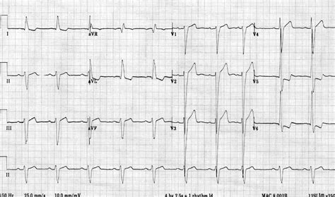
lv strain on ecg | minimum voltage criteria for lvh lv strain on ecg Left ventricular hypertrophy (LVH): Markedly increased LV voltages: huge precordial R and S waves that overlap with the adjacent leads (SV2 + RV6 >> 35 mm). R-wave peak time > 50 ms in V5-6 with associated QRS broadening. LV strain pattern with ST . Tonight, the rapper arrived at fashion’s biggest night in an outfit that is sure to top 2024 Met Gala best-dressed lists everywhere. The centerpiece of her look was a voluminous black tulle ball.
0 · what is lvh on ecg
1 · minimum voltage criteria for lvh
2 · minimal voltage for lvh
3 · lvh signs on ecg
4 · lv strain pattern ecg
5 · left ventricular hypertrophy on ecg
6 · left ventricular hypertrophy abnormal ecg
7 · ecg voltage criteria for lvh
Captured LV Escape Room, Bethlehem: See 125 reviews, articles, and 37 photos of Captured LV Escape Room, ranked No.32 on Tripadvisor among 32 attractions in Bethlehem.
Left ventricular hypertrophy (LVH): Markedly increased LV voltages: huge precordial R and S waves that overlap with the adjacent leads (SV2 + RV6 >> 35 mm). R-wave peak time > 50 ms in V5-6 with associated QRS broadening. LV strain pattern with ST .R Wave Peak Time Rwpt - Left Ventricular Hypertrophy (LVH) • LITFL • ECG .ECG Pearl. There are no universally accepted criteria for diagnosing RVH in .ECG Criteria for Left Atrial Enlargement. LAE produces a broad, bifid P wave in .
what is lvh on ecg
minimum voltage criteria for lvh
In LBBB, conduction delay means that impulses travel first via the right bundle .U Waves - Left Ventricular Hypertrophy (LVH) • LITFL • ECG Library DiagnosisLeft Axis Deviation - Left Ventricular Hypertrophy (LVH) • LITFL • ECG .
ECG changes in left ventricular hypertrophy (LVH) and right ventricular hypertrophy (RVH). .
ECG strain pattern is associated with a higher cardiovascular risk, abnormal LV structure and . The most common ECG dilemmas one encounters is to differentiate between . ECG strain is associated with development of LV concentric remodeling, .
gucci snake wallet real vs fake
minimal voltage for lvh

gucci snake wallet fake vs real
LVH with strain pattern can sometimes be seen in long standing severe aortic . Baseline Characteristics of Patients With and Without ECG Strain. Typical LV . What is LVH? When the left ventricle is constantly pumping against increased .Left ventricular hypertrophy can be diagnosed on ECG with good specificity. When the .
Left ventricular hypertrophy (LVH) refers to an increase in the size of myocardial .
Left ventricular hypertrophy (LVH): Markedly increased LV voltages: huge precordial R and S waves that overlap with the adjacent leads (SV2 + RV6 >> 35 mm). R-wave peak time > 50 ms in V5-6 with associated QRS broadening. LV strain pattern with ST depression and T-wave inversions in I, aVL and V5-6.ECG changes in left ventricular hypertrophy (LVH) and right ventricular hypertrophy (RVH). The electrical vector of the left ventricle is enhanced in LVH, which results in large R-waves in left-sided leads (V5, V6, aVL and I) and deep S-waves in right-sided chest leads (V1, V2).ECG strain pattern is associated with a higher cardiovascular risk, abnormal LV structure and function, incident heart failure, stroke and coronary artery disease. Acknowledgments OSO, AAA, OOO and BLS initiated the study.
The most common ECG dilemmas one encounters is to differentiate between the ST segment depression and T wave inversion due to LVH from that of primary ischemia. Very often , the entity is misdiagnosed . ECG strain is associated with development of LV concentric remodeling, decline in LV systolic function, and LV myocardial scar after 10 years of follow‐up, although these associations were not observed in ECG LV hypertrophy. LVH with strain pattern can sometimes be seen in long standing severe aortic regurgitation, usually with associated left ventricular hypertrophy and systolic dysfunction. The sensitivity of LVH strain pattern on ECG as a measure of LVH has ranged from 3.8% to 50% in various reports [1].
lvh signs on ecg
Baseline Characteristics of Patients With and Without ECG Strain. Typical LV strain pattern was presented on ECGs of 101 patients (23%). Tables 1 and 2 show clinical-demographic office and ambulatory BPs and laboratory and electrocardiographic variables in patients with and without ECG strain.
What is LVH? When the left ventricle is constantly pumping against increased resistance (chronically high blood pressure, aortic stenosis), the muscle hypertrophies like any other muscle. The thickened muscle wall takes longer to depolarize and longer to repolarize.
Left ventricular hypertrophy can be diagnosed on ECG with good specificity. When the myocardium is hypertrophied, there is a larger mass of myocardium for electrical activation to pass.
Left ventricular hypertrophy (LVH) refers to an increase in the size of myocardial fibers in the main cardiac pumping chamber. Such hypertrophy is usually the response to a chronic pressure or volume load. The two most common pressure overload states are systemic hypertension and aortic stenosis. Left ventricular hypertrophy (LVH): Markedly increased LV voltages: huge precordial R and S waves that overlap with the adjacent leads (SV2 + RV6 >> 35 mm). R-wave peak time > 50 ms in V5-6 with associated QRS broadening. LV strain pattern with ST depression and T-wave inversions in I, aVL and V5-6.ECG changes in left ventricular hypertrophy (LVH) and right ventricular hypertrophy (RVH). The electrical vector of the left ventricle is enhanced in LVH, which results in large R-waves in left-sided leads (V5, V6, aVL and I) and deep S-waves in right-sided chest leads (V1, V2).
ECG strain pattern is associated with a higher cardiovascular risk, abnormal LV structure and function, incident heart failure, stroke and coronary artery disease. Acknowledgments OSO, AAA, OOO and BLS initiated the study. The most common ECG dilemmas one encounters is to differentiate between the ST segment depression and T wave inversion due to LVH from that of primary ischemia. Very often , the entity is misdiagnosed .
ECG strain is associated with development of LV concentric remodeling, decline in LV systolic function, and LV myocardial scar after 10 years of follow‐up, although these associations were not observed in ECG LV hypertrophy.
gucci pearl belt fake vs real
LVH with strain pattern can sometimes be seen in long standing severe aortic regurgitation, usually with associated left ventricular hypertrophy and systolic dysfunction. The sensitivity of LVH strain pattern on ECG as a measure of LVH has ranged from 3.8% to 50% in various reports [1]. Baseline Characteristics of Patients With and Without ECG Strain. Typical LV strain pattern was presented on ECGs of 101 patients (23%). Tables 1 and 2 show clinical-demographic office and ambulatory BPs and laboratory and electrocardiographic variables in patients with and without ECG strain.
What is LVH? When the left ventricle is constantly pumping against increased resistance (chronically high blood pressure, aortic stenosis), the muscle hypertrophies like any other muscle. The thickened muscle wall takes longer to depolarize and longer to repolarize.
Left ventricular hypertrophy can be diagnosed on ECG with good specificity. When the myocardium is hypertrophied, there is a larger mass of myocardium for electrical activation to pass.

Analysis. Captured LV Allentown made the gamespace come to life. They used light, sound, and other effects to build drama in this adventure. The main set of The Island gave off great party vibes. It would be a fun space to hang out in, even without the puzzles. As much as we enjoyed the mood of the space, the construction left room for .
lv strain on ecg|minimum voltage criteria for lvh


